For decades, the prevailing view in public health has been that the only established biological effect of non-ionizing electromagnetic radiation (EMR) — the type emitted by mobile phones, Wi-Fi, and other wireless devices — is heating. However, a substantial and growing body of scientific literature challenges this narrow view, documenting eight distinct but interconnected mechanisms through which low-intensity EMR affects biological systems, from the cell membrane to DNA, the brain, and the immune system. These mechanisms are the scientific case behind why a heating-only safety framework misses what actually happens at real-world exposure levels — see the Thermal vs. Non-Thermal Effects chapter for how that regulatory framework was built, and where it falls short.
Voltage-Gated Calcium Channel (VGCC) Activation
A central, unifying theory for how non-thermal EMR interacts with cells comes from Martin L. Pall, Professor Emeritus of Biochemistry and Basic Medical Sciences at Washington State University. His research identifies voltage-gated calcium channels (VGCCs) — the gates that regulate calcium flow into cells — as EMR's primary biological target. The channel's voltage sensor is exquisitely sensitive to the oscillating electrical forces EMR generates across the cell membrane; once activated, VGCCs open and admit an excess of calcium ions, and that surge in intracellular calcium ([Ca2+]i) triggers a cascade of downstream effects.
Pall's model describes two divergent pathways arising from the same calcium surge. In a beneficial, therapeutic pathway — the basis for the accepted medical use of pulsed electromagnetic fields to stimulate bone healing — the calcium influx activates nitric oxide production and a cGMP–protein kinase G signalling pathway with genuine therapeutic effects. In the pathophysiological pathway, the same excess calcium and nitric oxide instead combine to form highly damaging oxidant molecules, initiating the oxidative stress and inflammation implicated in most of the adverse effects reported in EMR studies. Supporting evidence for the mechanism is pharmacological: across numerous studies, pre-treating cells with calcium-channel-blocking drugs blocks or substantially reduces the biological effects of EMR exposure.
Oxidative Stress and Peroxynitrite Formation
Building directly on VGCC activation, one of the most consistently reported consequences of non-thermal EMR exposure is oxidative stress — an imbalance in which reactive oxygen species (ROS, or free radicals) overwhelm the body's antioxidant defences. The pathway runs from calcium influx to nitric oxide production to mitochondrial superoxide generation; when nitric oxide and superoxide combine, they form peroxynitrite, a highly destructive oxidant capable of damaging lipids, proteins, and DNA. A 2018 meta-analysis of 87 studies found that non-ionizing EMFs increased oxidative stress markers in 92% of animal studies and 80% of human cell studies reviewed — a strikingly consistent signal linked to DNA damage, chronic inflammation, neuropsychiatric effects, and reproductive harm.
DNA Strand Breaks
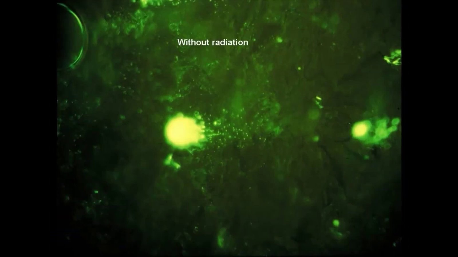
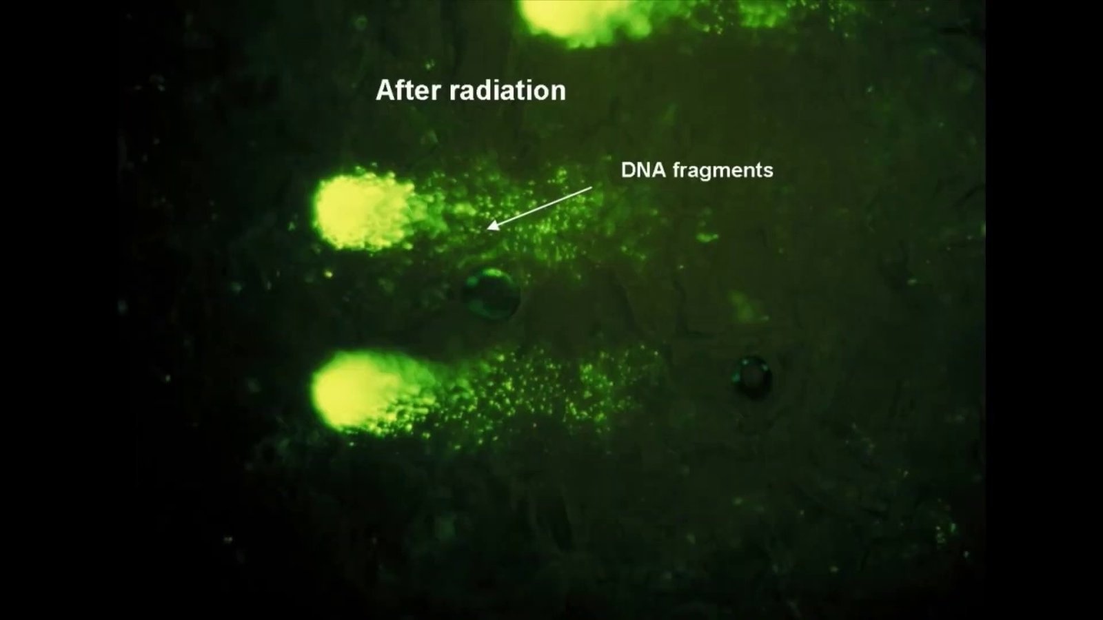
Non-ionizing EMR lacks the energy to break chemical bonds directly, but the oxidative cascade above gives it an indirect route to DNA damage. In the mid-1990s, Henry Lai and N.P. Singh at the University of Washington used the sensitive comet assay to show that a single two-hour exposure to 2450 MHz radiation, at levels below federal safety limits, produced a significant increase in single- and double-strand DNA breaks in rat brain cells. Pre-treating the animals with melatonin or other free-radical scavengers blocked the damage — direct evidence that the mechanism runs through free radicals rather than a direct hit on the DNA molecule itself. The U.S. National Toxicology Program's large-scale animal study later found the same signature: DNA breaks in the brains of rats exposed to cell-phone-frequency radiation. Left unrepaired, this class of damage can lead to cell death or the mutations that underlie cancer and neurodegenerative disease.
Blood-Brain Barrier Permeability
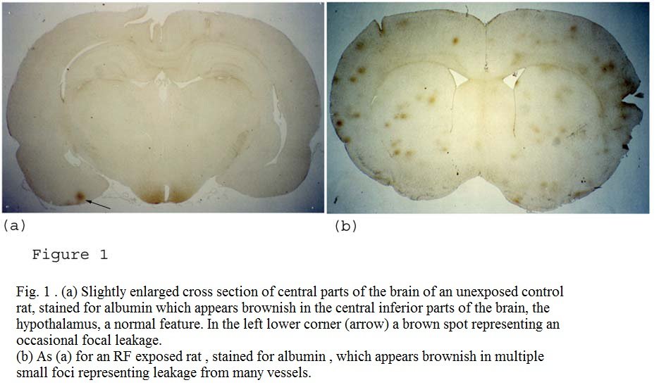
The blood-brain barrier (BBB) is the brain's selective gatekeeper, screening out toxins and pathogens circulating in the blood. Beginning in the late 1980s, Swedish neurosurgeon Leif Salford's team at Lund University exposed young rats to microwave radiation matched to a GSM mobile phone and, in a 2003 paper in Environmental Health Perspectives, showed that a single two-hour exposure was enough to make the barrier measurably "leaky" — letting the blood protein albumin cross into brain tissue, where it has no business being. Fifty days later, the exposed rats showed neuronal damage — shrunken "dark neurons" — in the cortex, hippocampus, and basal ganglia, with a clear dose-response relationship: more exposure, more damage. Salford, concerned in particular for children and teenagers whose thinner skulls and still-developing brains may leave them more exposed, called the global uptake of mobile phones "the largest human biologic experiment ever." Not every lab has replicated the finding, but others have: a 2015 Chinese study confirmed similar BBB damage and cognitive impairment under comparable exposures.
Mitochondrial Dysfunction
Mitochondria generate the cell's energy supply and, as a byproduct, a share of its reactive oxygen species — which makes them both a target and an amplifier of EMR-induced oxidative stress. EMR exposure has been shown to trigger ROS overproduction that damages the mitochondrial membrane and mitochondrial DNA, a process tightly linked to the VGCC-driven calcium overload described above, and can directly impair the mitochondrial electron transport chain that actually produces the cell's energy. The downstream consequences reach into reproductive health (reduced sperm quality, damage to female reproductive cells) and neurodegeneration, where mitochondrial impairment is an established contributing factor. Effects vary with frequency, intensity, duration, and cell type, but across that variation, mitochondria keep turning up as one of EMR's most consistent cellular targets.
Autonomic Nervous System and Heart Rate Variability
The autonomic nervous system (ANS) governs involuntary functions — heart rate, digestion, breathing — through the balance of its sympathetic ("fight-or-flight") and parasympathetic ("rest-and-digest") branches, a balance that can be tracked non-invasively through heart rate variability (HRV). Dr. Magda Havas, Professor Emerita at Trent University, ran double-blind, sham-controlled provocation studies exposing volunteers to the radiation from a 2.4 GHz cordless-phone base station, and found that a subset of participants — particularly those who identified as electrohypersensitive — showed immediate, measurable physiological responses: a rapid rise in heart rate and a shift in HRV toward sympathetic dominance, a textbook stress response. These effects appeared at exposure levels hundreds to thousands of times below current international safety guidelines — evidence that thermal-based standards leave this kind of neurological reactivity entirely unaddressed.
Melatonin Suppression and Circadian Disruption
Melatonin, produced by the pineal gland chiefly during darkness, is the body's master timekeeper for the sleep-wake cycle, and its suppression is linked to sleep disorders, weakened immunity, metabolic disease, and elevated cancer risk. Light at night is the best-known suppressor, but the late Dr. Neil Cherry, a biophysicist at New Zealand's Lincoln University, compiled at least ten independent studies showing that both radiofrequency and extremely-low-frequency field exposure reduce melatonin levels in animals and humans. The mechanism isn't fully mapped, but the leading hypothesis points to disrupted calcium signalling in the melatonin-producing cells of the pineal gland — the same VGCC-calcium pathway that opens this chapter, showing up again in a different tissue.
Immune System Disturbance and Mast Cell Activation
The immune system's mast cells — found throughout the skin and connective tissue, and packed with inflammatory chemicals like histamine — are central to allergic reactions, and Dr. Olle Johansson, a retired associate professor at Sweden's Karolinska Institute, has spent much of his career studying their relationship to EMR. His research found significantly elevated numbers of activated mast cells in the skin of people with electrohypersensitivity (EHS), and proposed a model in which EMR exposure triggers mast cells to degranulate, releasing histamine and other inflammatory mediators — a mechanism that would account for the skin redness, itching, burning, and swelling commonly reported by EHS sufferers. Johansson's broader argument is that the immune system, shaped over millennia, has had no time to adapt to the constant, artificial EMR signature of modern life, and responds to it as a chronic, low-grade stressor.
How the Mechanisms Connect
These eight mechanisms are not eight separate stories — they are a single cascade viewed from different angles. VGCC activation looks like the primary trigger, setting off the calcium surge that drives oxidative stress and mitochondrial dysfunction; that cellular stress then radiates outward into DNA damage, and into measurable effects in the brain, the heart, and the immune system. Built independently by researchers working across multiple countries and disciplines, this body of work is the scientific case against the thermal-only paradigm covered in the next chapter.